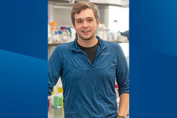
The National Institutes of Health’s (NIH) National Institute of Arthritis and Musculoskeletal and Skin Diseases has awarded Duke Anesthesiology’s Christopher Donnelly, PhD, DDS, (contact principal investigator) a three-year $5,734,530 grant for a translational research project titled, “Neural Architecture of the Murine and Human Temporomandibular Joint.”
This award was selected to be one of just five UC2 awards which make up the NIH’s Restoring Joint Health and Function to Reduce Pain (RE-JOIN) Consortium, which aims to define the innervation of the different articular and peri-articular tissues that collectively form the joint (including bone, cartilage, synovium, joint capsule, ligament, tendon, fascia and muscle), by sensory neurons that mediate the sensation of pain. The RE-JOIN Consortium is part of the broader NIH HEAL Initiative, an aggressive trans-agency effort to accelerate scientific solutions to treat pain and end the national opioid epidemic.
Donnelly will lead this award together with co-PIs, Joshua Emrick, DDS, PhD (University of Michigan School of Dentistry), and Dawen Cai, PhD (University of Michigan Medical School). Their multidisciplinary research team also consists of many other co-investigators across Duke, including Shad Smith, PhD (Anesthesiology); Aurelio Alonso, DDS, PhD (Anesthesiology); Zhicheng “Jason” Ji, PhD (Biostatistics and Bioinformatics); Yong Chen, PhD (Neurology); and Benjamin Hechler, DDS, MD (Plastic, Maxillofacial, and Oral Surgery).
This project led by Donnelly, Emrick and Cai, will comprehensively define the properties of sensory neurons that innervate tissues which form the temporomandibular joint. This work is significant because it will lead to an understanding of the neurological mechanisms that underlie chronic orofacial pain in patients with temporomandibular disorders (TMDs) and other orofacial pain conditions, thereby facilitating the development of new therapeutic approaches. Their project will also develop new technologies which will accelerate the study of TMDs and other joint pain conditions.
TMDs are the most common form of chronic orofacial pain, affecting five percent of adults in the United States. Despite substantial clinical and research interest in this area, progress to identify and target pathophysiological mechanisms underlying TMDs has been slow. This lackluster progress is owed in large part to our relatively primitive understanding of the basic neuroanatomical, molecular and physiological features of sensory afferents present within the temporomandibular joint (TMJ) tissues. The RE-JOIN Consortium seeks to address this knowledge gap through the formation of interdisciplinary teams which can define the innervation of articular and peri-articular tissues that collectively make up the jaw joint.
Donnelly, Emrick and Cai have partnered together to leverage their varied scientific expertise and define what they call the “neural architecture” of the sensory neurons which innervate the temporomandibular joint in animal models and in humans. “This is a large-scale, highly ambitious NIH initiative, and the RE-JOIN Consortium is expecting each research team to produce meaningful scientific discoveries that will lead to new pain therapies,” says Donnelly, assistant professor in anesthesiology. “One major reason that the NIH developed this sweeping program was to address a critical knowledge gap in our understanding of the basic features of joint innervation and joint pain. We knew our project had to be ambitious in its scope, and we wanted to understand the basic physiology of the sensory neurons which innervate the TMJ tissues from virtually every angle. Thus, our project will conduct experiments to define the cellular, molecular, and neurophysiological properties of orofacial sensory neurons in health and chronic pain states. Beyond accelerating our understanding of orofacial somatosensation, this project will reveal mechanisms of joint pain that can be used to develop new clinical interventions and pain therapies.”
To achieve their lofty research goals, the research team proposes to use MRI-guided delivery of retrograde dyes and viral tracers to achieve spatiotemporal precision, which they will combine with transcriptomics and advanced imaging approaches to map the molecular properties of peripheral sensory neurons which innervate distinct tissues within the murine TMJ in both steady-state and TMD conditions, using this information to build new intersectional genetic models to permit whole-TMJ mapping using lightsheet microscopy. In addition, using intersectional genetic approaches in conjunction with chemogenetics, in vivo Ca2+ imaging, and behavioral phenotyping, they will characterize the physiological/functional properties of TMJ-innervating sensory neurons, allowing them to identify neuronal subpopulations which contribute to chronic pain in TMD. To confirm that these findings bear relevance to human temporomandibular joint physiology, the team will establish a biobank of TMJ tissues from TMD-free healthy human donors and from a cohort of patients with chronic TMD pain, followed by quantitative analysis of peripheral afferent subtypes across TMJ tissues in each cohort. Lastly, they plan to build a free, user-friendly web-based platform to integrate the resulting transcriptomic, functional and macroscopic imaging datasets to permit widespread dissemination of these data. Overall, Donnelly states, “We anticipate these data will yield a working model of the sensory architecture of the temporomandibular joint tissues in mice and humans, including alterations in TMDs compared to steady-state conditions.”