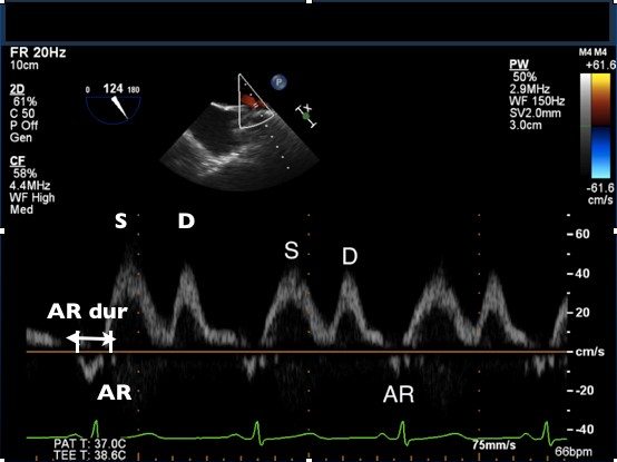
- Image the left pulmonary vein (or the right pulmonary vein)
- Align cursor with blood flow, use color flow Doppler for accuracy
- Place sample volume 1 cm into the pulmonary vein from the opening into the right atrium
- Use PW Doppler and record spectral doppler
- Using the preset measurements under ‘Analysis’ measure:
- Peak S velocity
- Peak D velocity
- Peak AR velocity
- AR wave duration