*The italicized text applies to patients undergoing mitral valve repair or replacement for mitral valve pathology or aortic valve replacement for aortic valve pathology
ME 4-chamber view with depth optimized for wall motion evaluation and strain (>50 Hz frame rate, 3 beats loop)
Color Doppler of MV, spectral Doppler with CW when significant regurgitation (measure peak velocity) or stenosis present (measure peak velocity, peak gradient and mean gradient)
Color Doppler of TV, spectral Doppler with CW when significant regurgitation or stenosis present
ME 5-chamber view
ME mitral commisural view
Color Doppler of MV
ME 2-chamber view with depth optimized for wall motion evaluation and strain (>50Hz frame rate, 3 beats loop)
Color Doppler of MV
ME left atrial appendage view
LA appendage (zoomed or less depth view, use X-plane), PW Doppler as appropriate (measure velocities)
ME LAX optimized for wall motion evaluation and strain (> 50 Hz frame rate, 3 beats loop)
Color Doppler of MV
Measure vena contracta if mitral regurgitation present
Proximal Isovelocity Surface Area if significant mitral regurgitation or stenosis present (measure PISA radius)
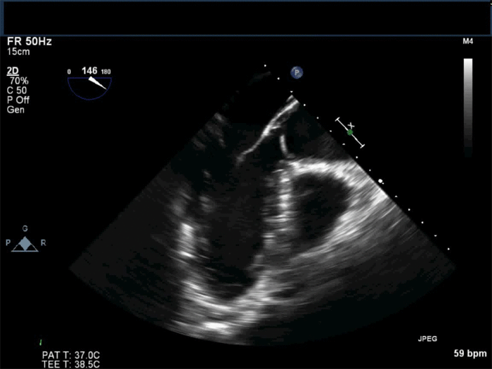
ME AoV LAX
Color Doppler of AoV
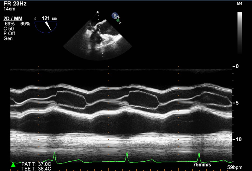
M-mode through the AoV
Measure vena contracta if aortic regurgitation present
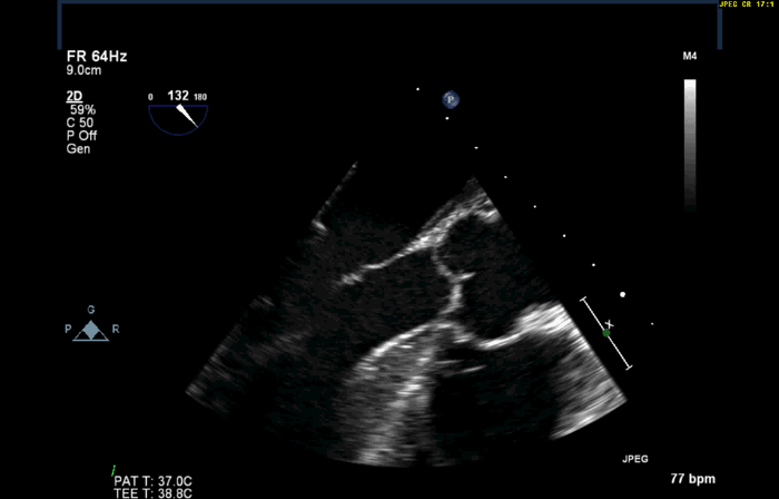
ME AoV SAX
Color Doppler AoV
ME Right ventricle inflow-outflow
Color Doppler of TV
Color Doppler of PV
ME bicaval view (proximal inferior & superior vena cava shown)
Color Doppler IAS (low Nyquist limit ≤40cm/s) to check for PFO/ASD
ME modified bicaval view
Color Doppler of TV, spectral Doppler with CW when significant regurgitation or stenosis present
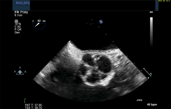
TG basal SAX (ensure good endocardial border visible. Use harmonics or a lower frequency if necessary. Ensure all walls visible if possible)
TG mid SAX
TG 2 chamber view
TG LAX
TG RV inflow view
TG RV inflow-outflow view
Deep TG 5-chamber view
Color Doppler of AoV
Spectral Doppler with CW if significant aortic regurgitation (measure pressure half-time) or stenosis (measure peak and mean gradients) present
Calculate AoV area using the continuity equation if aortic stenosis present
Descending aorta LAX and SAX (use X-plane)
UE aortic arch LAX and SAX

ME ascending aorta LAX and SAX
Diastology:
Quick guide to assessment of diastolic function - Cory Maxwell, MD
Renal arterial blood flow: spectral Doppler with PW
3D acquisition prebypass
3D Full Volume left ventricle
3D Full Volume right ventricle at 0 ME view with the RV tilted to be in the center of the image
3D Zoom mitral valve/3D Full Volume zoomed on mitral valve-surgeon's perspective
3D Full Volume with color Doppler of mitral valve if moderate or greater mitral valve regurgitation or stenosis
If aortic regurgitation or stenosis present:
3D Full Volume of the aortic valve at 120 ME view
3D Full Volume color acquisition of the aortic valve at 120 ME view
*The italicized text applies to patients undergoing mitral valve repair or replacement for mitral valve pathology or aortic valve replacement for aortic valve pathology
ME 4-chamber view with depth optimized for wall motion evaluation and strain (>50 Hz frame rate, 3 beats loop)
Color Doppler of MV
Color Doppler of TV
ME mitral commissural view
Color Doppler of TV
ME 2-chamber view with depth optimized for wall motion evaluation and strain (>50Hz frame rate, 3 beats loop)
Color Doppler of MV
ME LAX optimized for wall motion evaluation and strain (> 50 Hz frame rate, 3 beats loop)
Color Doppler of MV
Measure vena contracta if mitral regurgitation present
ME AoV LAX
Color Doppler of AoV
M-mode through the AoV
ME AoV SAX
Color Doppler AoV
ME Right ventricle inflow-outflow
Color Doppler of TV
Color Doppler of PV
ME bicaval view (proximal inferior & superior vena cava shown)
Color Doppler IAS (low Nyquist limit ≤40cm/s) to check for PFO/ASD
ME modified bicaval view
Color Doppler of TV, spectral Doppler with CW when significant regurgitation or stenosis present
TG basal SAX (ensure good endocardial border visible. Use harmonics or a lower frequency if necessary. Ensure all walls visible if possible)
TG mid SAX
TG 2 chamber view
TG LAX
Deep TG 5-chamber view
Descending aorta LAX and SAX (use X-plane)
UE aortic arch LAX and SAX
ME ascending aorta LAX and SAX
Record gradients through the new prosthetic valve or valve repair
Diastology:
Transmitral flow
Pulmonary vein flow
Tissue Doppler imaging
Propagation velocity
Renal arterial blood flow: spectral Doppler with PW (measure peak systolic flow and minimal diastolic flow)
3D acquisition postbypass
3D Full Volume left ventricle
3D Full Volume right ventricle at 0 ME view with the RV tilted to be in the center of the image
3D Zoom mitral valve/3D Full Volume zoomed on mitral valve-surgeon's perspective after mitral valve repair or replacement
3D full volume with color acquisition of the mitral valve after mitral valve replacement and repair
3D Full Volume color acquisition of the aortic valve at 120 ME view
Additions and exceptions from the postbypass protocol for left ventricular assist devices:
Visualize the inflow cannula in the ME views with and without color
Visualize the outflow cannula in the ascending aorta views. Record velocities with CW Doppler
Do not perform diastology
Do not assess left ventricular wall motion and ejection fraction
Additions to the postbypass protocol for right ventricular assist devices:
Visualize the inflow cannula in the right atrium
Visualize the outflow cannula in the pulmonary artery (ME Asc Ao SAX and/or UE aortic arch SAX). Record velocities with CW Doppler.
Postbypass perform a thorough examination of the transplanted heart following the “Prebypass protocol” above.
Additions to the postbypass protocol
Image right and left pulmonary veins with and without color
Record velocities in the right and left pulmonary veins using PW Doppler
Image main pulmonary artery and right pulmonary artery with and without color (ME Asc Ao SAX and/or UE Aortic Arch SAX)
PRE-PROCEDURE At least 2-beat loops, clear ECG signal
3D acquisition
3D Full Volume left ventricle
3D Zoom mitral valve/3D Full Volume zoomed on mitral valve-surgeon's perspective
3D Full Volume with color Doppler of mitral valve
3D Full Volume of the aortic valve at 120 ME view
3D Full Volume color acquisition of the aortic valve at 120 ME view
2D acquisition
ME 4-chamber view with depth optimized for wall motion evaluation (FR >50 Hz)
Color Doppler of MV, spectral Doppler with CW when significant regurgitation (measure peak velocity) or stenosis present (measure peak velocity, peak gradient and mean gradient)
Color Doppler of TV, spectral Doppler with CW when significant regurgitation or stenosis present
ME bicommisural view
Color Doppler of MV
ME 2-chamber view with depth optimized for wall motion evaluation (FR> 50Hz))
Color Doppler of MV
LA appendage (zoomed, use X-plane), PW Doppler as appropriate (measure velocities)
ME LAX optimized for wall motion evaluation (FR >50Hz)
Color Doppler of MV
Measure vena contracta if mitral regurgitation present
Measure PISA radius if significant mitral regurgitation or stenosis present
ME LAX of the AoV
Color Doppler of AoV
Measure vena contracta if aortic regurgitation present
Measure LVOT, annulus, sinuses of Valsalva, STJ, ascending aorta diameters
ME SAX of the AoV
Color Doppler AoV
ME Right ventricle inflow-outflow
Color Doppler of TV
Color Doppler of PV
ME bicaval view (proximal inferior & superior vena cava shown)
Color Doppler IAS (low Nyquist limit ≤40cm/s) to check for PFO/ASD
TG basal SAX (ensure good endocardial border visible. Use harmonics or a lower frequency if necessary. Ensure all walls visible if possible)
TG mid SAX
TG 2 chamber view
TG LAX
Deep TG view
Color Doppler of AoV
Spectral Doppler with CW if significant aortic regurgitation (measure pressure half-time) or stenosis (measure peak and mean gradients) present
Spectral Doppler with PW across the LVOT
Calculate AoV area using the continuity equation if aortic stenosis present
Diastology:
Transmitral flow
Pulmonary vein flow
Tissue Doppler imaging
Propagation velocity
Renal arterial blood flow: spectral Doppler with PW (measure peak systolic velocity and minimal diastolic velocity)
Descending aorta LAX and SAX (use X-plane)
Aortic arch LAX and SAX
Ascending aorta LAX and SAX
POST-PROCEDURE
Please evaluate of the new prosthetic aortic valve in the following views:
Record prosthetic aortic valve loops/images after the transvalvular wire has been removed.
ME SAX
Color Doppler of AoV
ME LAX
Color Doppler of AoV
TG LAX
Color of AoV
Spectral Doppler with CW across the prosthetic valve
Deep TG
Color Doppler of AoV
Spectral Doppler with CW across the prosthetic valve
Spectral Doppler with PW across the LVOT
Other images
ME 4-chamber view with depth optimized for wall motion evaluation (FR >50 Hz FR)
Color Doppler of MV
Color Doppler of TV
ME 2-chamber view with depth optimized for wall motion evaluation (FR >50Hz)
Color Doppler of MV
ME LAX with depth optimized for wall motion evaluation (FR> 50 Hz)
Color Doppler of MV
ME Right ventricle inflow-outflow
Color Doppler of TV
Color Doppler of PV
TG basal SAX (ensure good endocardial border visible. Use harmonics or a lower frequency if necessary. Ensure all walls visible if possible)
TG mid SAX
TG 2 chamber view
TG LAX
Diastology:
Transmitral flow
Pulmonary vein flow
Tissue Doppler imaging
Descending aorta LAX and SAX (use X-plane)
Aortic arch LAX and SAX
Ascending aorta LAX and SAX
3D acquisition
3D Zoom mitral valve/3D Full Volume zoomed on mitral valve-surgeon's perspective
3D Full Volume with color Doppler of mitral valve
3D Full Volume of the aortic valve at 120 ME view
3D Full Volume color acquisition of the aortic valve at 120 ME view