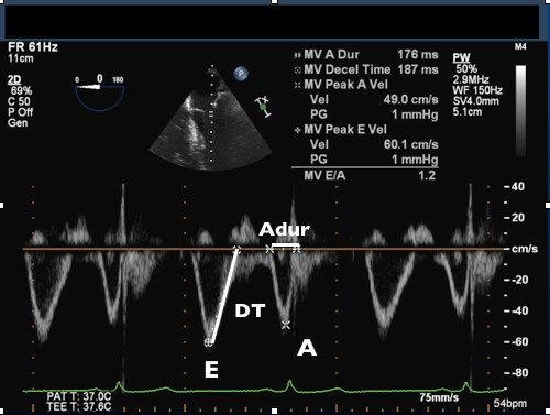
- Obtain a mid-esophageal 4-chamber view
- Align cursor with blood flow through the mitral valve (use color flow Doppler if needed)
- Place sample volume at the level of the mitral valve tips in diastole
- Use pulse-wave Doppler
- Using the preset measurements available under ‘Analysis’ measure:
- Peak E velocity
- Peak A velocity
- Deceleration time of the E wave
- A wave duration
