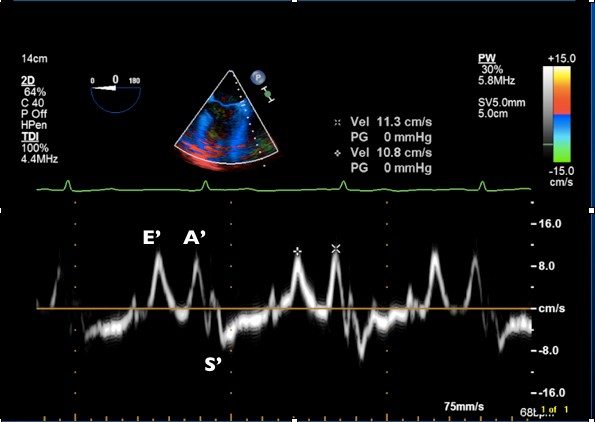
- Obtain a mid-esophageal 4-chamber view
- Activate the Tissue Doppler Imaging function of the TEE machine
- Place sample volume at the level of the lateral mitral annulus
- Use pulse-wave Doppler
- Using the preset measurements available under ‘Analysis’ measure:
- Peak E’ velocity
- Peak A’ velocity