- Obtain a transgastric SAX mid-papillary
- Turn the probe 90-270° and advance 4-5 cm
- As you turn you may visualize the left kidney (central echogenic pelvis and hypoechogenic peripheral cortex)
- If by turning to the left you visualize the descending aorta, turn the probe back to the right about 90° and find the left kidney
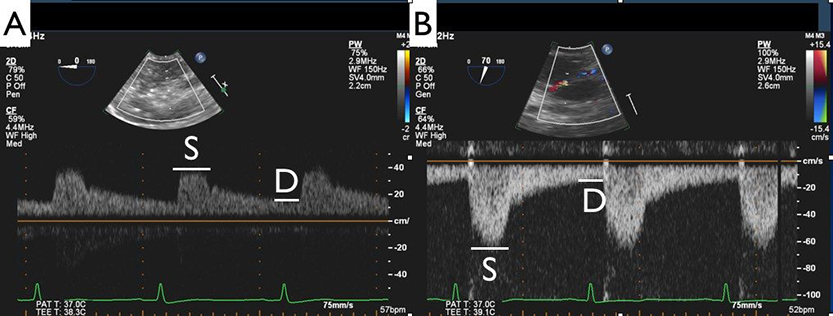
- Zoom in the left kidney, apply color flow Doppler (decrease the scale and increase color flow gain to visualize blood flow through the kidney)
- Apply PW Doppler
- Measure peak systolic velocity (S) and minimum diastolic velocity (D)
- Renal resistivity index (RRI) = (S-D)/S
- Perfect alignment of the PW Doppler beam with the blood flow is not necessary as we are not interested in velocities but in the ratio of these velocities.
