- Obtain a mid-esophageal 4-chamber view
- Align color flow with the transmitral flow, change the size of the box to include both the mitral annulus and the left ventricular apex
- Change the color map baseline towards blue to a velocity of 20 cm/sec
- Align the Doppler beam with the blood flow through the mitral valve
- Activate M-mode
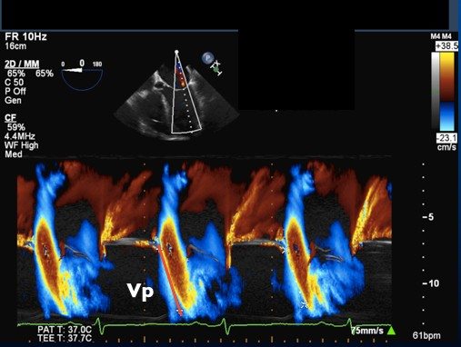
- You should obtain a waveform pattern as in the image below
- Measure the slope of the first aliasing velocity from the mitral annulus to 4 cm into the LV.